
Figure 1.
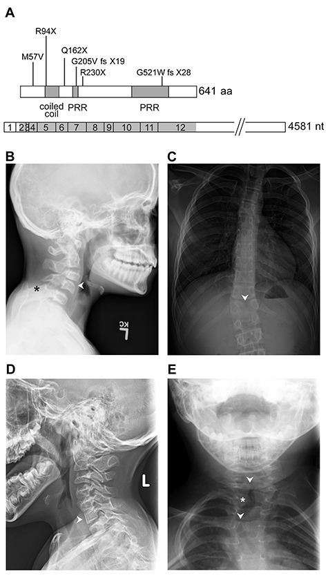
WBP11 variants and vertebral segmentation defects in selected individuals. (A) Schematic representation of the WBP11 open reading frame and cDNA (bottom) with variants and protein structure (top). PRR, proline-rich region. (B) Patient 1A has C3-C4 (arrowhead), C6-C7 fusion/block vertebrae (not shown) and bilateral omovertebral bones from C5 (asterisk). (C) Patient 1B has T12 butterfly vertebrae (arrowhead) and congenital scoliosis. Patient 2 has C6-C7 fusion (D, arrowhead) and T2 left hemifusion (E, asterisk) and T1, T4 butterfly vertebrae (E, arrowheads).