
Figure 6.
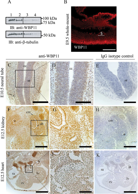
WBP11 is ubiquitously expressed in mouse embryos. (A) Immunoblot showing WBP11 (lane 1) and WBP11-FLAG (lane 2) overexpression in C2C12 mouse cell line and endogenous WBP11 expression in C2C12 (lane 3) and E9.5 embryo lysate (lane 4). Beta-tubulin was used as a loading control. (B) Whole-mount immunofluorescence staining of an E9.5 mouse embryo with anti-WBP11 antibody. Anti-WBP11 reactivity was detected with donkey anti-rabbit RRX. (C–K) Transverse sections of paraffin embedded mouse embryos at different stages labeled with anti-WBP11 antibody (left two columns) or IgG isotype control (right column) followed by detection with anti-rabbit HRP and DAB staining. (C–E) Sections through the neural tube at E10.5. (F–H) Sections through the kidney at E12.5. (I–K) Sections through the heart at E12.5. All paraffin sections were counterstained with hematoxylin, dorsal is to the top and ventral to the bottom. s, somite; nt, neural tube; cc, central canal; cm, condensed mesenchyme; ub, uretric bud; rv, right ventricle; lv, left ventricle; ra, right atrium; la, left atrium. Scale bars: 500 μm (I, K), 300 μm (C, E, F, H), 100 μm (B, D, G, J).