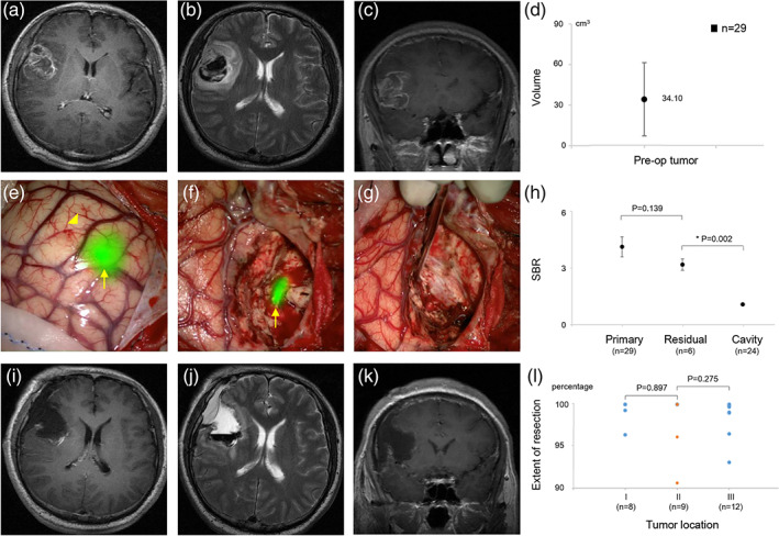
FIGURE 2.

Preoperative, intraoperative, and postoperative imaging data of fluorescence‐guided surgical resection of a glioblastoma multiforme (GBM) with IRDye800‐BBN. (a–c) Reprehensively preoperative enhancing brain magnetic resonance imaging (MRI) of a right frontal GBM. (d) Preoperative contrast‐enhanced tumor volume. (e) Immediately after opening the dura, the tumor (arrow) showed an obvious fluorescence; the normal cortical area and superficial vein (arrowhead) showed no fluorescence. (f) The residual tumor (arrow) showed an obvious fluorescence. (g) The tumor cavity did not exhibit any obvious fluorescence, indicating a complete resection. (h) The signal‐to‐background ratio (SBR) values of the primary tumor, residual tumor, and tumor cavity. (i–k) Postoperative MRI confirmed a lack of residual tumor. (l) Extent of resection in different tumor locations
