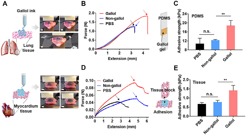
Fig. 7.
On-tissue 3D printing and adhesion of the gallol ECM hydrogel ink at 1:2 mass ratio and 6 wt%. (A) Schematics and images of printing of the gallol ink (containing blue food coloring dye) on porcine lung (top) and myocardium (bottom) tissues. Scale bars of 4 mm. (B) Force-extension curves and (C) quantification of adhesive strengths for the tensile testing of gallol hydrogels used to adhere PDMS substrates. The inset schematic shows the sample design and the arrows within the curves indicate the detachment of the materials from the PDMS substrates. (D) Force-extension curves and (E) quantification of adhesive strengths for the tensile testing of printed gallol ECM hydrogel lattices used to adhere myocardial tissues. The inset schematic corresponds to the experimental design for testing and the arrows within the curves indicate the detachment of the lattices from the myocardial substrates. Sidak test in conjunction with one-way ANOVA, n.s. (not significant) and **p < 0.01.

