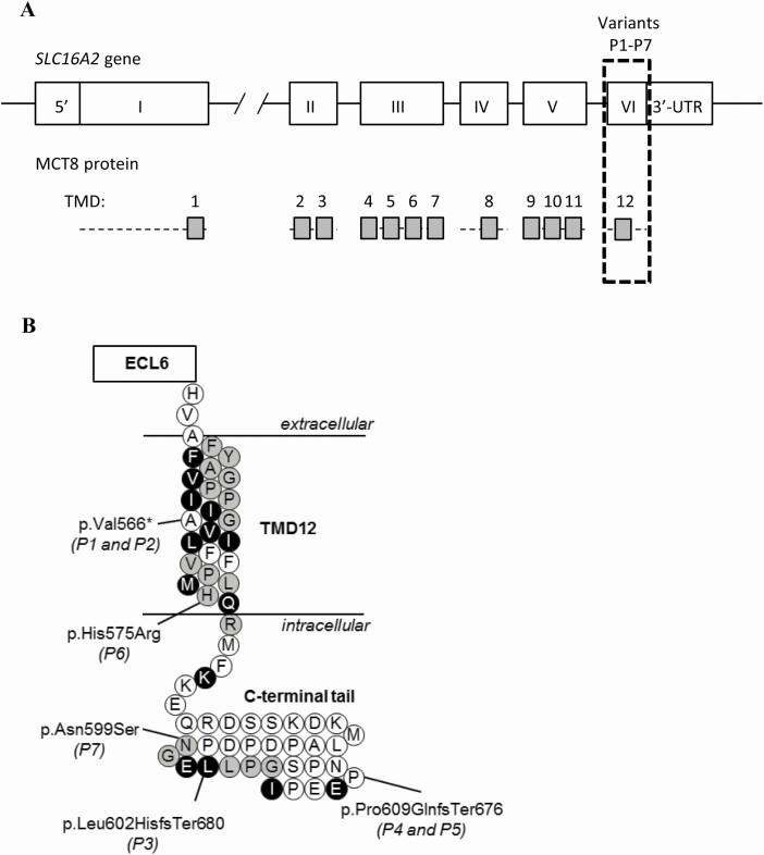
Figure 1.
(A) Schematic representation of the SLC16A2 gene and MCT8 protein. The transmembrane domains (TMDs) are displayed as grey boxes and aligned to their coding exons. All variants identified in this study locate to exon 6 (boxed with dashed lines). (B) Schematic representation of TMD12 and the intracellular C-terminal tail. The locations of the identified variants are indicated, according to the reference sequence of the long isoform (NM_006517.3, NP_006508.1). Strongly conserved residues are colored black, whereas those with strongly similar properties across species are colored grey (see Supplemental Fig. 2 for detailed alignment (24)). Abbreviations: ECL, extracellular loop.

