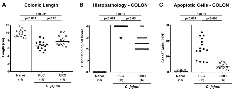
Figure 3.
Macroscopic and microscopic inflammatory outcomes upon urolithin-A application to C. jejuni infected IL-10−/− mice. Following C. jejuni strain 81-176 infection on days 0 and 1, microbiota depleted IL-10−/− mice were treated with urolithin-A (URO, open circles) or placebo (PLC, closed circles) via the drinking water starting on d2 post infection. On d6, (A) the colonic lengths were measured and (B) the histopathological changes in large intestinal ex vivo biopsies quantified using standardized histopathology scores. (C) Furthermore, the average numbers of apoptotic colonic epithelial cells (positive for cleaved caspase3, Casp3+) were determined microscopically from six high power fields (HPF, 400 × magnification) per mouse applying in situ immunohistochemical analysis of large intestinal paraffin sections. Naive mice were used as negative controls (open diamonds). Medians (black bars), significance levels (p values) calculated by the ANOVA test with Tukey post-correction or by the Kruskal–Wallis test and Dunn’s post-correction and the total numbers of included animals (in parentheses) are given. Data were pooled from three independent experiments.

