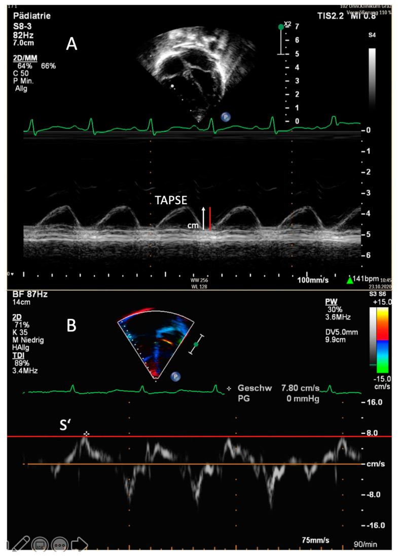
Figure 2.
Transthoracic imaging. (A) Apical four-chamber view. Determination of the tricuspid annular plane systolic excursion (TAPSE) in a 1-year-old patient with idiopathic pulmonary arterial hypertension in M-mode. The red line demonstrates a TAPSE value that is abnormally low in this patient (1.1 cm; z-score −3). (B) Apical four-chamber view. Right, ventricular tissue Doppler imaging (TDI) with the PW curser placed at the lateral tricuspid annulus in an 8-year-old patient with pulmonary arterial hypertension associated with congenital heart disease (PAH-CHD). In this patient, the values of the tricuspid peak systolic excursion (S’) are reduced, depicted as the red line.

