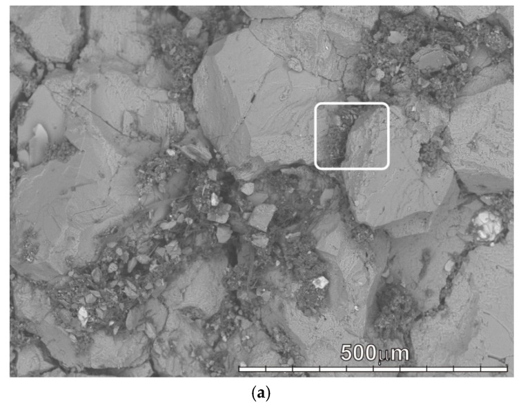
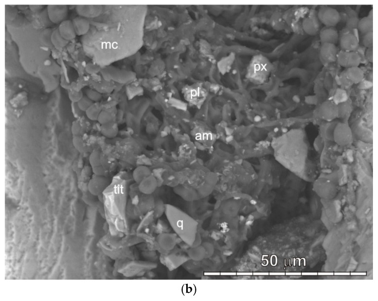
Figure 2.
SEM-image of a biofilm with a predominance of microscopic fungi on the surface of a homogeneous calcite marble: (a)—microcolonies of fungi around calcite grains (the white frame shows the area shown in figure b); (b)—grains of various silicate minerals among fungal hyphae and rounded cells (in the region shown in Figure 2a): quartz (q), plagioclase (pl), mica (mc), pyroxene (px), Fe—titanite (tit), Pb—amphibole (am).


