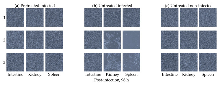
Figure 5.
Intestinal epithelial cells (IECs), intestine, kidney, and spleen tissues cultured from olive flounder (Paralichthys olivaceus). (a) Replicates of intestine, kidney, and spleen homogenates cultured in EPC cells from the orally B. subtilis-pretreated olive flounder at a concentration of 106 CFU/mL examined 96 h after the VHSV infection. Note the absence of cytopathic effects (CPE) in all the tissues examined. (b) Replicates of intestine, kidney, and spleen homogenates cultured in EPCs from the B. subtilis-untreated olive flounder examined 96 h after the VHSV infection. Note the presence of CPE in two cases of the EPC cells inoculated with spleen and kidney samples and the absence of CPE in all intestine replicates. (c) Replicates of intestine, kidney, and spleen homogenates cultured in EPC cells from B. subtilis-untreated olive flounder that were not infected by VHSV examined after 96 h. Note the absence of CPE in all the tissues examined. All images were visualized by phase-contrast microscopy at 100× magnification.

