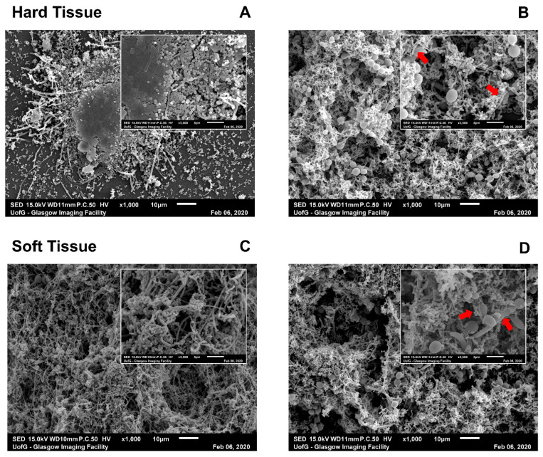
Figure 3.
SEM images of hard tissue (HT) and soft tissue (ST) biofilms with or without C. albicans. Biofilms were processed after maturation and viewed on a JEOL JSM-6400 scanning electron microscope. Images were captured at ×1000 magnification (main image) and ×3500 magnification (inset). HT (A,B) and ST biofilms (C,D) were imaged without (A,C) and with C. albicans (B,D). Red arrows in insets of Candida-containing biofilms indicated visible attachment of bacterial cells with fungal hyphae or yeast cells. Images chosen were representative of duplicate samples.

