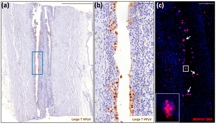
Figure 2.
Detection of polyomavirus large-T antigen and BKPyV DNA in the ureter of a native kidney removed from patient 9 due to the presence of a clear cell renal cell carcinoma. (a) Scan of a tissue section stained for large-T antigen (scale bar: 500 μm). (b) Region corresponding to the blue square highlighted in panel (a) (scale bar: 100 μm). This section was counterstained with hematoxylin to visualize cell nuclei. (c) Serial section stained for BKPyV genome by fluorescent in situ hybridization (FISH) (red) (inset: magnification of the white square). The white arrows indicate FISH-BKPyV-positive nuclei. This section was counterstained with 4′,6-diamidino-2-phenylindole (DAPI) (blue) to visualize cell nuclei (scale bar: 100 μm).

