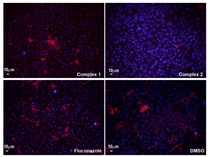
Figure 6.
The adhesion of fluorescent C. albicans SC5314-RFP on A549 cells in the presence of complexes 1 and 2, and fluconazole (Olympus BX51, Applied Imaging Corp., San Jose, CA, United States, under 20× magnification; scale bar representing 10 µm). DAPI (2-(4-amidinophenyl)-6-indolecarbamidine dihydrochloride) stained nuclei appear in blue, while fluorescent red is from labeled C. albicans cells.

