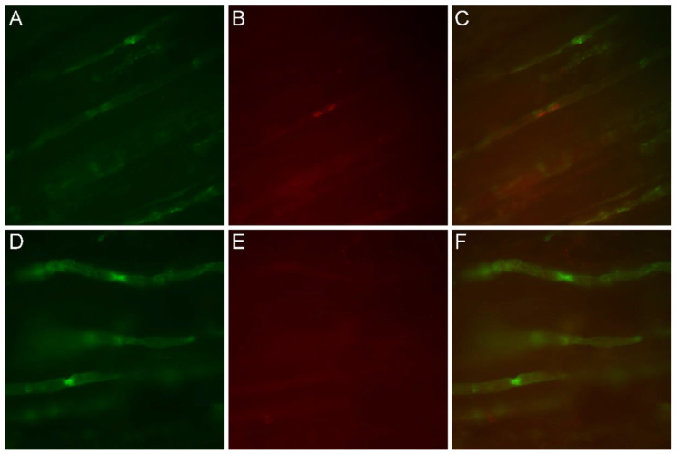
Figure 4.
Immunofluorescence study on nerve longitudinal sections, comparing patient III-8 (D–F) to a normal control (A–C). Original magnification 40×. (A,D) Green immunofluorescence following reaction with anti-MAG antibodies. (B,E) Red immunofluorescence using anti-Cx32 antibodies. (C,F) Merged images. In the control nerve sample, anti-MAG and anti-Cx32 antibodies colocalized at the paranodes (C), whereas in the patient, Cx32 signal was lacking (E) and the sole anti-MAG immunofluorescence was observed in merged images (F).

