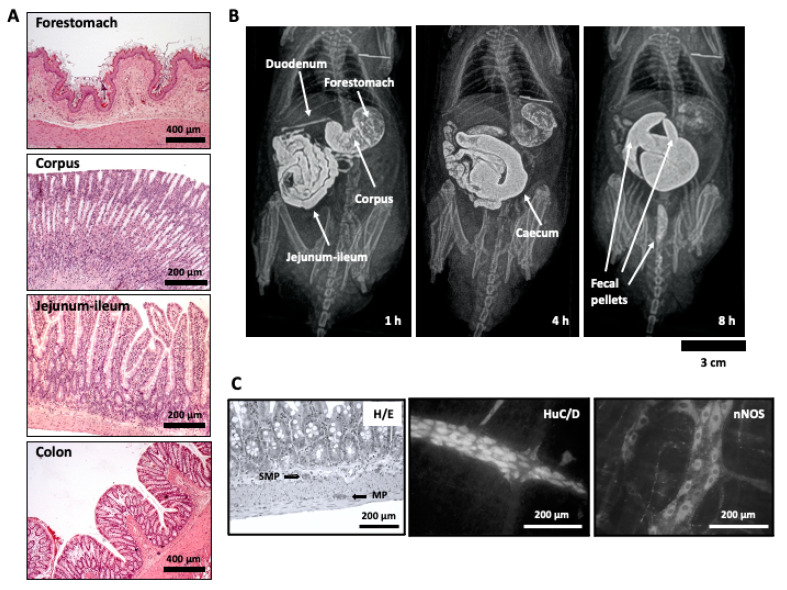
Figure 1.
(A) Histological appearance of the wall of forestomach, corpus, jejunum-ileum (the longest part of the small intestine), and colon. (B) Organs of the rat gastrointestinal tract visualized by radiographic methods at different time points after intragastric barium administration in a conscious rat. Since rats do not vomit, barium can only progress towards the anus: 1 h after contrast, the two parts of the rat stomach (forestomach and corpus) can be distinguished, as well as the duodenum and the jejunum-ileum; 4 h after contrast, the stomach and small intestine can still be partially seen but now the cecum is filled with contrast; 8 h after contrast, the stomach and small intestine are barely seen, but the cecum is well filled with contrast and some fecal pellets are present within the colon. (C) Microscopic images showing the appearance of the enteric nervous system: left, location of the submucous (SMP) and the myenteric plexuses (MP) within the rat ileal wall in histological sections, stained with hematoxylin/eosin (H/E); middle and right, whole-mount or “sheet-like” preparations (from guinea pig ileum), obtained after dissecting away mucosa, submucosa, and circular muscle, leaving behind only the longitudinal muscle layer with the myenteric plexus attached; whole-mount preparations were processed immunohistochemically to show all the neurons with the pan-neuronal marker HuC/D (middle), or the specific subpopulation of neurons immunoreactive to neuronal nitric oxide synthase (nNOS), for which both somata and nerve fibers, but not nuclei, can be distinguished (right).

