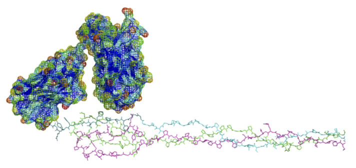
Figure 8.
Preliminary molecular simulation of docking between the scFv antibody and the α2Ct epitope of rat collagen. Lower figure: C-terminus of the rat type I collagen model; α1(I) chains, purple and green; α2(I) chain, cyan, α2Ct epitope, dark cyan. Upper figure: scFv antibody with highly water accessible “exposed” regions (mesh surface) color-scaled towards the red end of the spectrum; green is intermediate and blue is not exposed. Antibody subunits show potentially complementary binding interactions at α2Ct epitope (see text).

