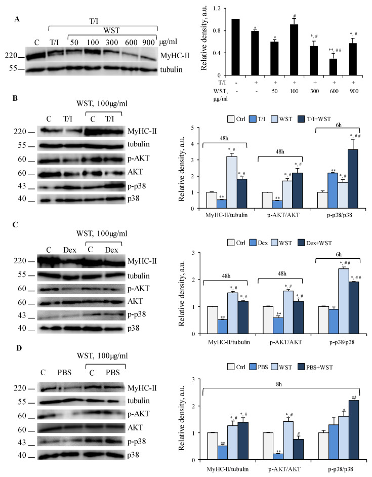
Figure 4.
(A) C2C12 myotubes were treated with TNFα (tumor necrosis factor α, 20 ng/mL)/IFNγ (interferon γ, 100 U/mL) (T/I) in the absence or presence of different doses of WST for 48 h and myosin heavy chain (MyHC)-II expression were analyzed by WB (western blotting). (B–D) WST was tested on myotubes untreated or treated with T/I (B) or dexamethasone (Dex, 1 µM) (C), or starved with PBS (phosphate buffered saline) (D) for indicated time-points. MyHC-II, p-AKT (phospho-protein kinase B), AKT, p-p38 MAPK (phosphor-p38 mitogen-activated protein kinase) and p38 MAPK expression were analyzed by WB. Tubulin was used as a loading control (A–D). Reported are representative images and the relative densities with respect to tubulin or total form of phosphorylated protein (A–D). Results are means ± SD (standard deviation) (A–D). Statistical analysis was conducted using t-test * p < 0.05, ** p < 0.01 significantly different from untreated control; # p < 0.05 and ## p < 0.01 significantly different from T/I (A,B), Dex (C) or PBS (D).

