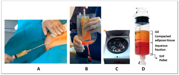
Figure 1.
Diagram of the Hy-Tissue stromal vascular fraction (SVF) process. (A) Lipoaspiration; (B) mechanic disaggregation of adipose tissue using the double bag; (C) centrifugation, (D) phase separation: From up to down, it is possible to distinguish the following layers: Oil, condensed fat, aqueous fraction, and a bottom pellet with the SVF product.

