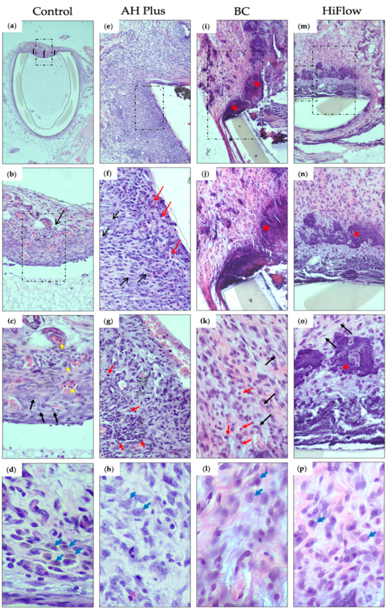
Figure 1.
Hematoxylin and eosin histological sections of the interface tissue-sealer 8 days after subcutaneous implantation (dashed boxes mark the view of the subsequent image): (a) control group, evidencing the thin fibrous capsule at the interface between the host tissue and the polyethylene tube (demarcation with dotted lines), measured in 3 points (represented by the black lines) (40× magnification); (b) the fibrous capsule and mild inflammatory reaction (score 1) (black arrow) at the interface tissue-sealer (200×); (c) high magnification showing in detail the cellular population consisting of polymorphonuclear leukocytes (neutrophils) (yellow arrows) and fibroblasts (black arrows) (400×); (d) macrophages (blue arrows) (800×); (e) AH Plus group, showing granulation tissue surrounding the polyethylene tube (demarcation with dotted lines) (100×); (f) high magnification evidencing small congested neo-capillaries (red arrows), fibroblasts (black arrows) and moderate inflammatory reaction (score 2) (200×); (g) high magnification, showing the inflammation with mainly lymphocytes (red arrows) and neutrophils (400× hematoxylin and eosin (H&E)). (h) Macrophage infiltration (blue arrows) (800×); (i) TotalFill BC Sealer (BC) group, showing a fibrous capsule with calcification (bluish deposits) (red asterisk) (100×). (j) Higher magnification showing fibroblasts and some inflammatory cells (score 1) and the calcified area in more detail (red asterisk) (200×). (k) Fibroblasts (fusiform cells) (black arrows) in a stroma with some collagen fibrils and some lymphocytes (red arrow), plasma cells and rare neutrophils (400×). (l) Macrophage infiltration (blue arrows) (800×); (m) HiFlow group, revealing a fibrous capsule with calcification next to the polyethylene tube (100×); (n) higher magnification to observe fibroblasts, and inflammatory cells (score 1) next to the calcified area (red asterisk) (200×); (o) calcified area (red asterisk), with fibroblasts (fusiform cells) (black arrows) in an edematous and low collagenous stroma with lymphocytes (400×); (p) macrophage infiltration (800×). (n = 8).

