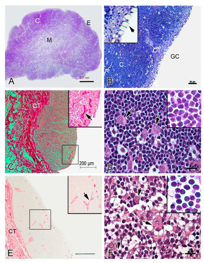
Figure 2.
Histological organization of the thymus in rainbow trout. (A). Thymus section of rainbow trout. Epithelium (E), cortex (C) and medulla (M) are shown. (B). High magnification of E and C. Gill cavity is indicated (GC). Thymus parenchyma is divided by connective tissue (CT, trabeculae). The inset shows the single layer of epithelium (E) with mucous-like cells arrow). (C). Shows a broad distribution of collagen (in red) in the CT. The inset shows blood vessels are positive for picrosirius staining (arrow). (D). High magnification of the C showing thymocytes and larger cells (15 µm diameter approximate, arrows). The inset shows thymocytes with a central spherical nucleus and scant cytoplasm. (E). Shows a broad distribution of elastic fibers in the CT (in brownish red). The inset shows blood vessels are positive for orcein staining (arrow). (F). High magnification of the M showing large cells (arrows); thymocytes (inset) with a similar morphology to those in the C. A, B, D and F—Giemsa histochemical staining. (C)—Picrosirius histochemical staining. (E)—Orcein histochemical staining. Note the distinction between the cortex (dark staining) and the medulla (pale staining).

