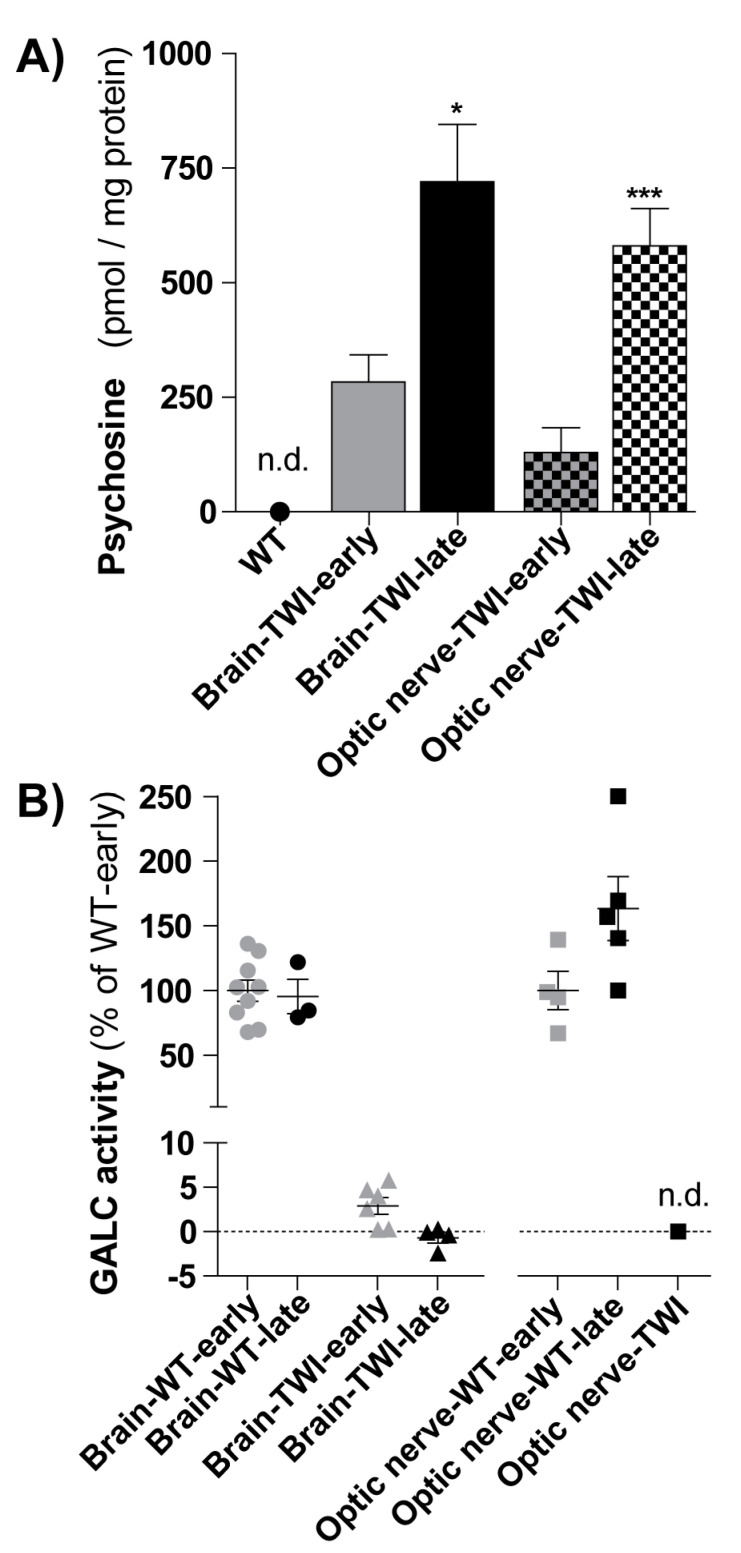
Figure 3.
(A) Psychosine (PSY) quantification. PSY content was measured in brains (full-filled color columns) and optic nerves (squared columns) of TWI mice in the early stage of disease (early: PND20-28, n = 7) and in the late stage of the disease (late: PND30-38, n = 12 for brain and n = 8 for optic nerve). */*** p < 0.05/0.001 early vs. late, Student’s t-test. PSY was not detectable (n.d.) in WT mice, in both districts. Data = mean ± SEM. (B) Galactosylceramidase (GALC) quantification. GALC activity (reported in percentage in respect to the respective WT-early) was measured in brains (left; circles and triangles) and optic nerves (right; squares) of WT and TWI mice in the early stage of disease (early, in grey: PND20-21 for brains, PDN18 for optic nerves) and in the late stage of the disease (late, in black: PND30-38). GALC was not detectable (n.d.) in the optic nerves of TWI mice. Each point represents a mouse; data= mean ± SEM.

