Figure 3.
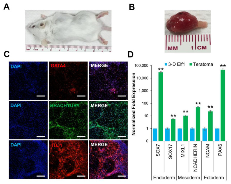
3-D cultured naïve ESCs produced teratomas in SCID-beige mice. (A) Tumor growth was observed in all mice injected with 3-D grown Elf1 cells (n = 3). (B) Explanted tumor at 4 weeks showed well-circumscribed lobular morphology consistent with teratoma formation. (C) Confocal images (10×) depict the presence of GATA4, BRACHYURY, and TUJ1 protein expression representing endoderm, mesoderm, and ectoderm, respectively. All scale bars represent 100 μm. (D) Gene expression analysis by qRT–PCR showed that teratomas expressed SOX7, and SOX17 (endoderm), MIXL1, and N-CADHERIN (mesoderm), and NCAM, and PAX6 (ectoderm). Results are expressed as the fold expression ± SEM normalized to reference genes HMBS and GAPDH (** p ≤ 0.01).

