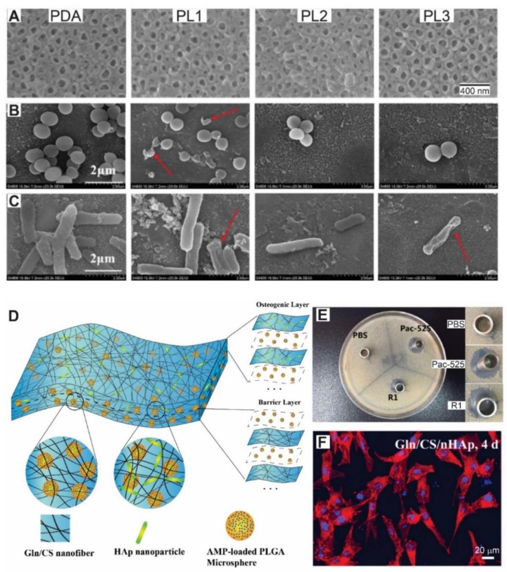
Figure 4.
(A) Top view SEM images of titanium dioxide nanotubes coated with poly-dopamine (PDA), PDA grafted with on layer of ε-Polylysine (PL1), PDA coated with two layers of ε-Polylysine and one layer of gum arabic (PL2) and PDA coated with three layers of ε-Polylysine and two layer of gum arabic (PL3). Morphology of S. aureus (B) and E. coli (C) adhered to each material examined by SEM. Cracked bacteria appear on the surface (red arrows). An antiadhesive effect can be seen with the increase in the number of layers (PL2 and PL3) along with the contact-kill effect. (D) Schematic diagram of the fabrication and structure of the composite membrane by Layer-by-Layer electrospinning of gelatin (Gln) and chitosan (CS), and alternatively electrospraying of antimicrobial peptide (Pac-525)-loaded poly lactic-co-glycolic acid microspheres. The osteogenic layer also includes hydroxyapatite (Hap) nanoparticles. (E) Typical inhibition zones of S. aureus induced by PBS (negative control), PAC-525 (positive control) solution and the supernatant solution after one week of incubation with the composite membranes described in (D). (F) Microscopy images of rat bone marrow mesenchymal stem cells stained for actyn cytoskeleton and cell nucleus 4 days after seeding on the osteogenic layer. A, B and C adapted with permission from [115]. Elsevier, 2020. (D–F) adapted from [116], MDPI, 2018.

