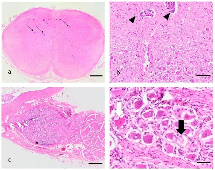
Figure 1.
(a–d): Histopathological changes in the medulla oblongata (a,b) and the spinal ganglia (c,d) of the diseased alpaca, hematoxylin and eosin stain. (a): Note the presence of multifocal prominent perivascular cuffs (arrows), bar 2 mm. (b): Perivascular cuffs consist predominantly of lymphocytes (black arrow heads), bar 100 µm. (c): Lymphocytes also infiltrate the spinal ganglia (asterisk), bar 500 µm. (d): Some neurons are shrunken and pale (black arrow) or display hypereosinophilia with pyknosis and chromatolysis (white arrow) (neuronal degeneration and necrosis), bar 50 µm.

