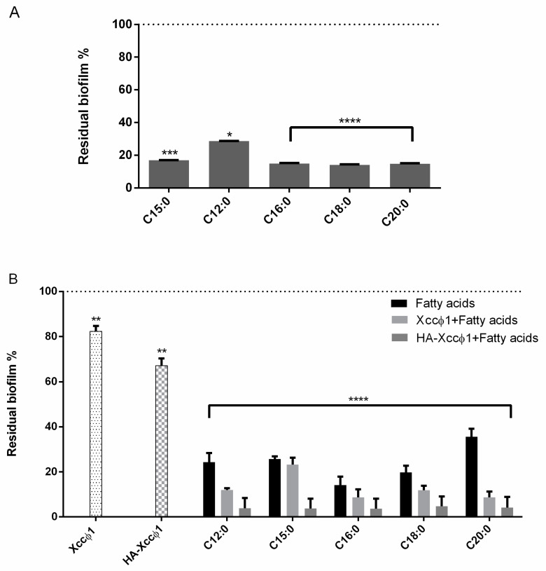
Figure 1.
(A) Analysis of the effect of different fatty acids (60 µg/mL) on Xcc mature biofilm structure after 72 h of incubation at 25 °C and 8 h of treatment. The data are reported as percentages of residual biofilm. Each value is the mean ± SD of three independent experiments. Statistical analysis was performed with the absorbance compared to the untreated control and considered statistically significant when p < 0.05 (* p < 0.05, ** p < 0.01, *** p < 0.001, **** p < 0.0001) according to two-way ANOVA multiple comparisons. (B) Analysis of the effect of all the acids fatty acids (60 µg/mL) with Xccφ1 or Xccφ1 plus HA on Xcc biofilm structure using crystal violet assay after 72 h of incubation at 25 °C and 6 h of treatment. The data are reported as percentages of residual biofilm. Each value is the mean ± SD of three independent experiments. Statistical analysis was performed with the absorbance compared to the untreated control and considered statistically significant when p < 0.05 (* p < 0.05, ** p < 0.01, *** p < 0.001, **** p < 0.0001) according to two-way ANOVA multiple comparisons.

