Figure 4.
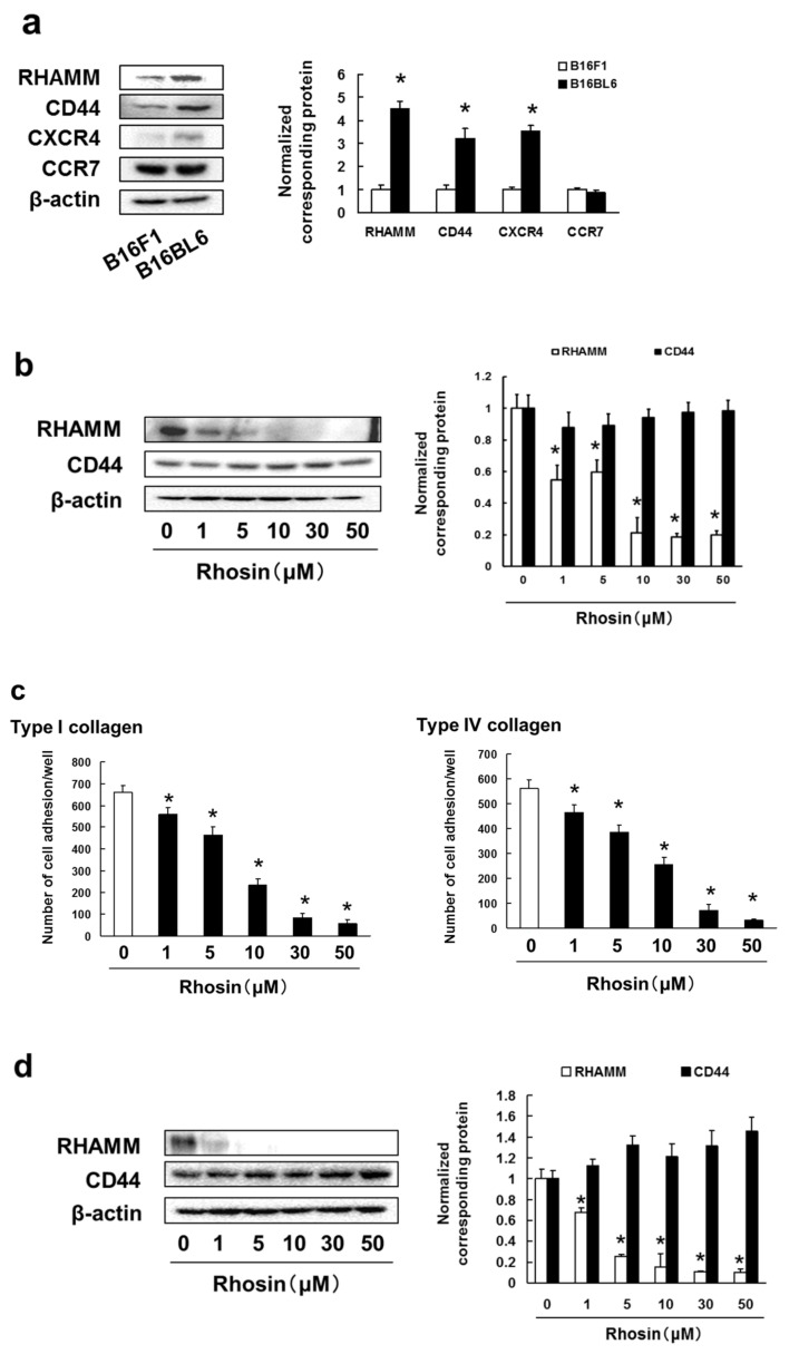
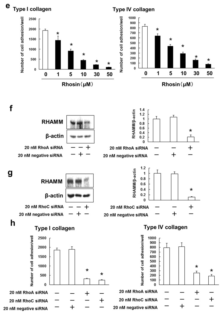
High metastatic potential cell lines of B16BL6 cells enhanced expression of RHAMM, CD44 and CXCR4: (a) Images of Western blots for the RHAMM, CD44, CXCR4, CCR7 and β-actin (internal standard), and quantification of the amounts of RHAMM, CD44, CXCR4 and CCR7 after normalization to the amounts of corresponding protein. The results are representative of 4 independent experiments. * p < 0.01 vs. B16F1 cells (ANOVA with Dunnett’s test); (b) B16BL6 cells were treated with rhosin at indicated concentration for 3 days. Images of Western blots for the RHAMM, CD44 and β-actin (internal standard), and quantification of the amounts of RHAMM and CD44 after normalization to the amounts of corresponding protein. The results are representative of 4 independent experiments. * p < 0.01 vs. controls (ANOVA with Dunnett’s test); (c) B16BL6 cells (1 × 10⁴ cells), which had been treated with rhosin for 3 days, were incubated with type I collagen- or type IV collagen-coated plates for 30 min at 37 °C in an atmosphere containing 5% CO2. The results are representative of 5 independent experiments. * p < 0.01, as compared to the controls (0.1% DMSO-treated) (ANOVA with Dunnett’s test); (d) 4T1 cells were treated with rhosin at indicated concentration for 3 days. Images of Western blots for the RHAMM, CD44 and β-actin (internal standard), and quantification of the amounts of RHAMM and CD44 after normalization to the amounts of corresponding protein. The results are representative of 4 independent experiments. * p < 0.01 vs. controls (ANOVA with Dunnett’s test); (e) 4T1 cells (1 × 10⁴ cells), which had been treated with rhosin for 3 days, were incubated with type I collagen- or type IV collagen-coated plates for 30 min at 37 °C in an atmosphere containing 5% CO2. The results are representative of 5 independent experiments. * p < 0.01, as compared to the controls (0.1% DMSO-treated) (ANOVA with Dunnett’s test); (f,g) B16BL6 cells were treated with negative siRNA, (f) RhoA siRNA, or (g) RhoC siRNA at indicated concentration for 3 days. Images of Western blots for the RHAMM and β-actin (internal standard), and quantification of the amounts of RHAMM after normalization to the amounts of β-actin. The results are representative of 4 independent experiments. * p < 0.01 vs. controls (ANOVA with Dunnett’s test); (h) B16BL6 cells (1 × 10⁴ cells), which had been treated with negative siRNA, RhoA siRNA, or RhoC siRNA for 3 days, were incubated with type I collagen- or type IV collagen-coated plates for 30 min at 37 °C in an atmosphere containing 5% CO2. The results are representative of 5 independent experiments. * p < 0.01, as compared to the controls (0.1% DMSO-treated) (ANOVA with Dunnett’s test).


