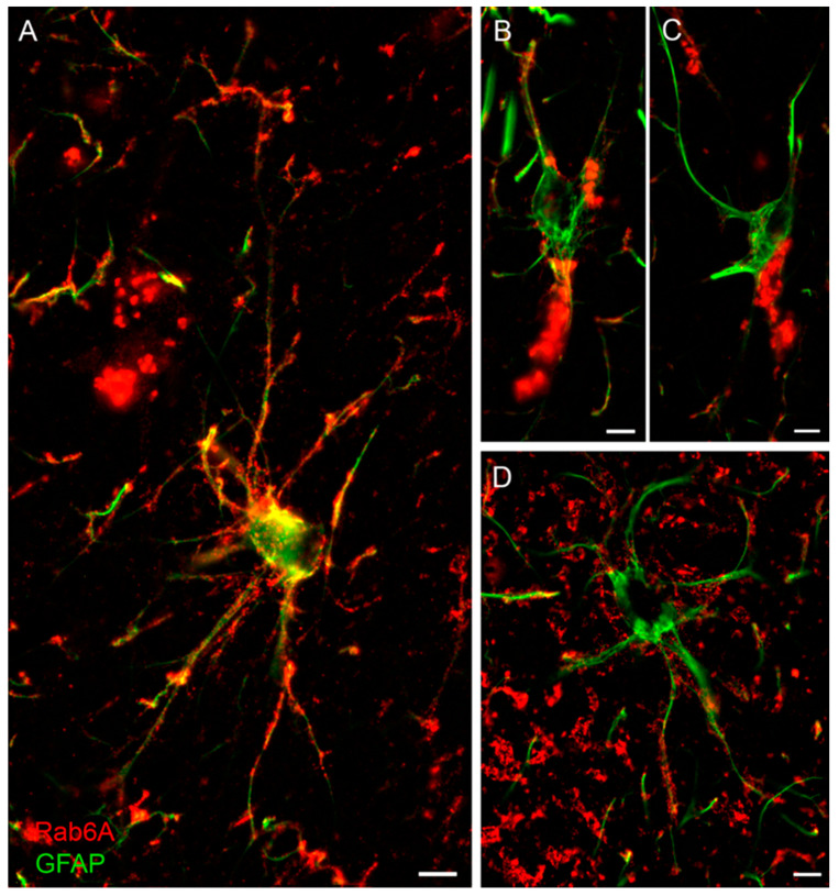
Figure 7.
Rab6A in human GFAP+ astrocytes. (A–D) Examples from three cases. All of the preselected human non-reactive GFAP+ astrocytes and most of the reactive GFAP+ astrocytes were Rab6A+ (for quantification, see Figure S11E). Note variability of cellular Rab6A staining with pronounced label in the distal processes (A,D) or predominant staining in the perinuclear region (B,C). Scales: 10 µm (A,D), 3 µm (B), 5 µm (C).

