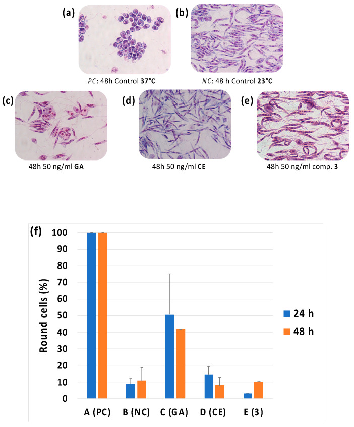
Figure 6.
Optical microscope photography (magnification ×1000) of Giemsa-stained smears of L. infantum promastigotes incubated for 48 h (a) at 37 °C to induce a reversible differentiation, positive control (PC); (b) at 23 °C, negative control (NC); (c) at 23 °C in the presence of 50 ng/mL of GA, (d) CE and (e) compound 3. In (f) the percentage of round amastigote-like cells after 24 h and 48 h of incubation is reported in the different conditions.

