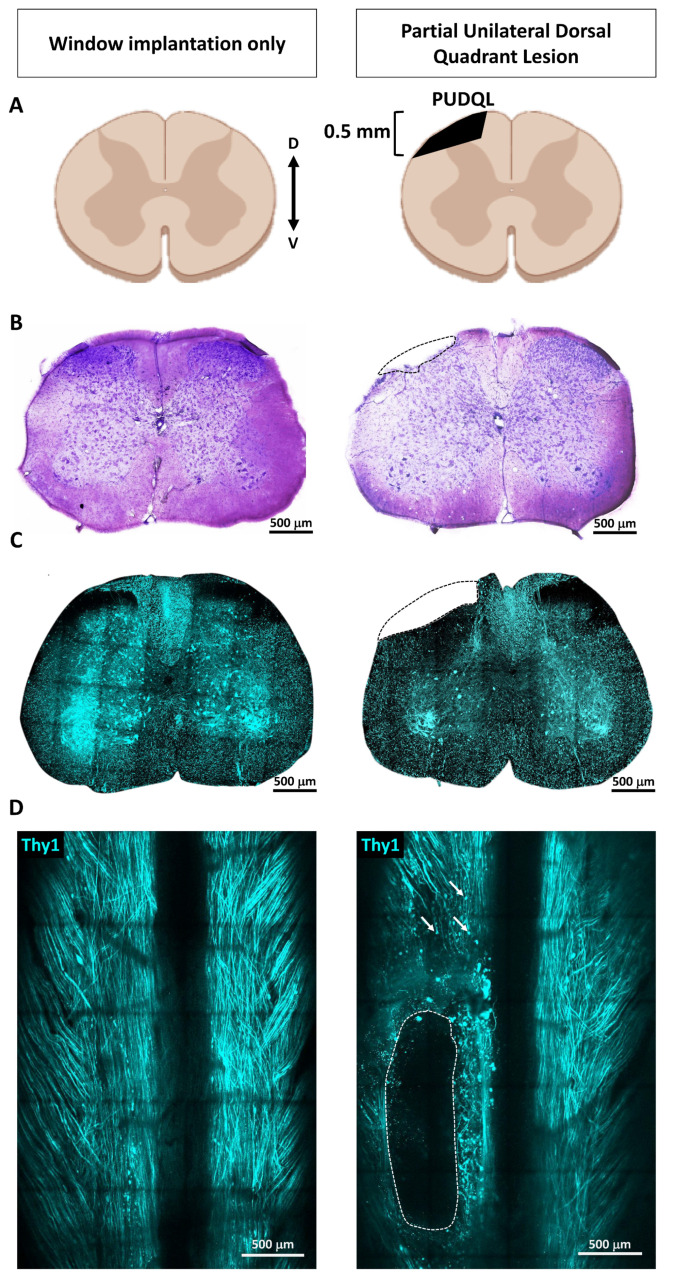
Figure 1.
Spinal cord partial unilateral dorsal quadrant lesion (PUDQL) in Thy1-CFP mice. (A) Schematic view of the PUDQL (shadowed area in black). (B) Cresyl violet staining of transversal spinal cord section collected three days (D3) after window implantation or after PUDQL followed by window implantation. (C) Two-photon (2P) fluorescence images of spinal slices consecutive to the one of B). (D) Dorsal view of the same Thy1-CFP spinal cord imaged through the implanted window showing the primary lesion area (dotted lines) and examples of subsequent Wallerian degeneration of axons (arrows) on D3. D: dorsal; V: ventral.

