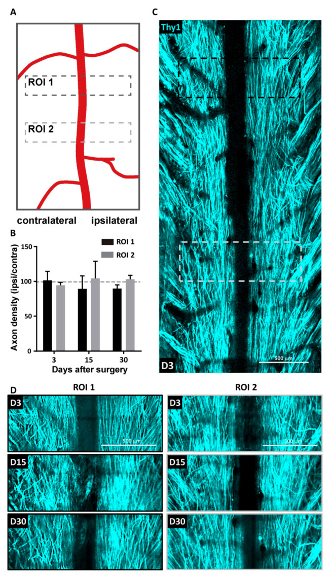
Figure 2.
Absence of axonal degeneration following spinal cord window implantation. (A) Regions of interest (ROI) used to monitor the stability of axonal pattern over 30 days in unlesioned animals (B) Evolution of the axonal density over time. Density on one side was normalized to the contralateral side. Data are shown as mean ± SEM (n = 5). (C) Representative maximal intensity projection 2P images obtained in vivo three days (D3) after window implantation. (D) Zoom in images obtained 3, 14 and 30 days after window implantation for the same ROI1 (black) and ROI2 (gray) highlighted in (C).

