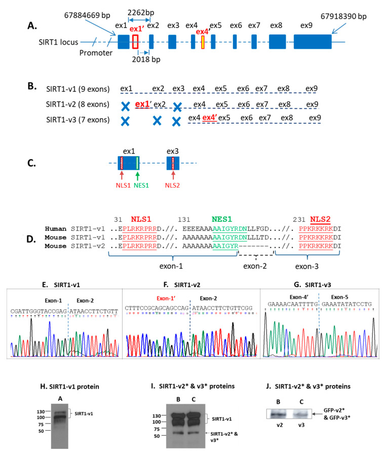
Figure 3.
Bioinformatic and experimental analysis of human SIRT1-v1, v2, and v3 isoforms. (A). SIRT1 gene locus and genomic DNA landscape. (B). SIRT1-v1 isoform contained nine exons, whereas SIRT1-v2 contained eight exons, in which the first exon had not been previously reported and it was named exon-1′ (ex1′), SIRT1-v3 isoform contained seven exons. The Exon-4′ (ex4′) was immediately behind exon-4 and it had not been previously reported. (C). Exon-1 (ex1) and exon-3 (ex3) contained nuclear localization signal sequences. (D). Sequence analysis of human SIRT1-v1 in comparison with both mouse SIRT1-v1 and v2. During alternative splicing, mouse SIRT1-v2 lost exon-2, but retained exon-1 and exon-3 that have NLS. Therefore, both mouse SIRT1-v1 and v2 isoforms are likely to be localized in the nucleus. (E–G). The sequencing chromatogram image of exon-junctions that were observed in SIRT1-v1 (Figure 3E), SIRT1-v2 (Figure 3F), and SIRT1-v3 (Figure 3G). (H–J). SIRT1 isoform protein expression in cell lysate samples that were transfected with plasmid constructs SIRT1-v1 (A), SIRT1-v2 (B), and SIRT1-v3 (C). (I). SIRT1-v2 and v3 protein expression. (J). GFP-SIRT1-v2 and –v3 fusion proteins were detected by anti-GFP antibody. * The molecular weight of SIRT1-v2 is 50.5 kDa, and SIRT1-v3 is 49.25 kDa, respectively.

