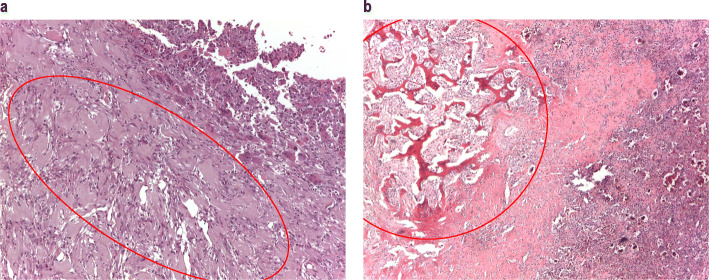
Fig. 4.
Histologic features of malignancy in GCTB. a Primary malignant GCTB, pre-denosumab: proliferation of ovoid to spindle bland-appearing cells, with scattered reactive multinucleated osteoclast-like giant cells (top right of image), consistent with GCTB, juxtaposed to a proliferation of atypical spindle and pleomorphic cells, growing in fascicles, consistent with undifferentiated pleomorphic sarcoma (red circle). b Secondary malignant GCTB, pre-denosumab, (recurrence in 2008): histological features consistent with GCTB (bottom right of image), juxtaposed to a proliferation of atypical spindle cells, infiltrating in between the host bony trabeculae, consistent with high-grade undifferentiated spindle cell sarcoma (red circle)

