
FIGURE 1.
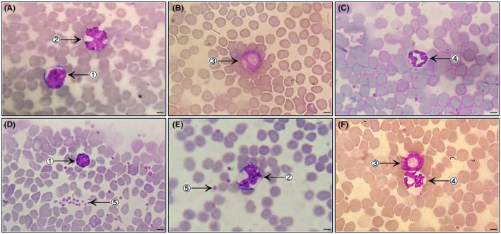
Photomicrographs of bloods smears (Diff‐Quick staining; 1000×; bar = 500 µm) from healthy mice: Swiss female (A and B) and male (C); BALB/c male (D and E) and female (F). The smears show red blood cells present in large quantities with uniform distribution, and evidence of leukocytes: lymphocyte (1), monocyte (2), eosinophil (3), neutrophil (4) and platelets (5)