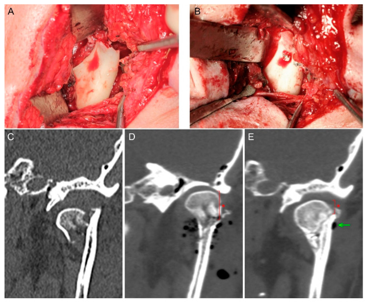
Figure 3.
Mandibular head fracture fixation. The phenomenon of reduced mandibular ramus height appeared (asterisk). (A) Fracture site. (B) Osteosynthesis by two magnesium 1.7 mm × 14 mm screws. (C) Pre-op scan—the mandibular head is dislocated downward which shortens the mandibular ramus. (D) Fixation by compressive headless screws; visible gas bubbles are the air introduced into the wound during open reduction; height of mandible head is marked by an asterisk. (E) 6-month post-op follow-up—fixed bone remodeling, remnants of the produced hydrogen gas (arrow), shortening of the mandibular ramus as a result of the proximal fragment (mandible head) down-shifting along the fissure of the fracture (asterisk).

