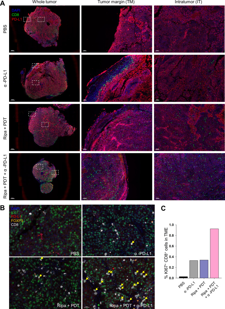
Figure 7.
Population and location of immune cell subsets in tumor using multiplex IHC. (A) Exact location and density of CD8+ cells (green) and PD-L1+ cells (red) in the TME were investigated using multiplex IHC. (B) Tumor tissues were stained with Ki67 (green), PD-L1 (red), FOXP3 (orange), CD8 (white) and nuclei was costained with DAPI (blue). Yellow arrows represents the proliferative CD8+ T cells. (C) Percentage of proliferative CD8+ (Ki67+CD8+) cells. IHC, immunohistochemistry; TME, tumor microenvironment.

