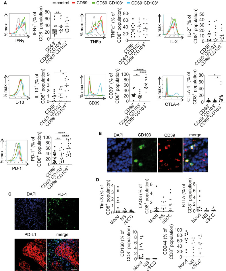
Figure 5.
CD8+CD69+CD103+ TRMs in cSCC are associated with increased expression of IL-10, CD39, CTLA-4 and PD-1. (A) Representative FACS histograms and accompanying graphs showing expression of IFNγ (n=14 tumors), TNFα (n=11 tumors), IL-2 (n=11 tumors), IL-10 (n=10 tumors), CD39 (n=14 tumors), CTLA-4 (n=14 tumors) and PD-1 (n=11 tumors) by the tumor-infiltrating CD8+CD69−, CD8+CD69+CD103− and CD8+CD69+CD103+ T-cell populations. Expression of cytokines was determined following stimulation in vitro with PMA and ionomycin for 5 hours. Horizontal bars=means, *p<0.05, **p<0.01, ****p<0.0001. (B) Representative immunofluorescence microscopy images of cSCC stained for CD103 and CD39. (C) Representative confocal microscopy images of cSCC showing PD-1 and PD-L1 expression. (D) Percentages of CD8+ T cells from blood, normal skin (NS) and cSCC expressing Tim3 (n=13 tumors), LAG3 (n=14 tumors), BTLA (n=11 tumors), CD160 (n=13 tumors) and CD244 (n=12 tumors). (B and C) Scale bars=50 µm. cSCC, cutaneous squamous cell carcinoma; TRMs, resident memory T cells.

