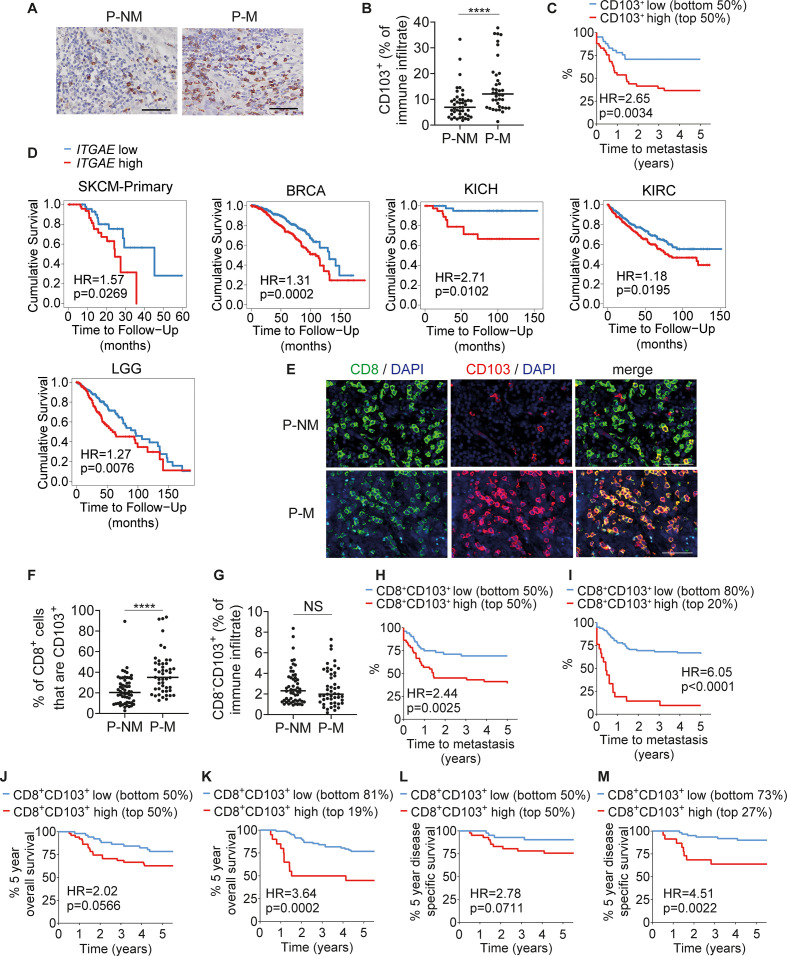
Figure 6.
CD103+ TRMs in primary cSCCs are associated with development of metastasis. (A) Representative immunohistochemistry images of cSCC stained for CD103. (B) Percentages of tumor-infiltrating immune cells that are CD103+ in primary cSCCs that did not metastasize (P-NM, n=44) and primary cSCCs that metastasized (P-M, n=38). (C) Kaplan-Meier plot showing days to metastasis for the cSCCs in (B) stratified by low (<9.25% of immune infiltrate, n=41) and high (≥9.25% of immune infiltrate, n=41) expression of CD103. (D) Survival analyzes by ITGAE expression based on TCGA data in primary cutaneous melanoma (SKCM-Primary, n=103), breast carcinoma (BRCA, n=1100), kidney chromophobe cancer (KICH, n=66), kidney renal clear cell carcinoma (KIRC, n=533) and lower grade glioma (LGG, n=516). (E) Representative immunofluorescence microscopy images of cSCCs stained for CD8 and CD103. (F) Percentages of CD8+ cells that are CD103+ in P-NM (n=56) and P-M (n=47) cSCCs. (G) Percentages of immune cells that are CD8−CD103+ in P-NM and P-M cSCCs. (H, I) Time to metastasis for the cSCCs in (F) split into low and high CD103+ cell frequencies as a percentage of the CD8+ cell population divided at (H) the median (low ≤26.04% of CD8+ population, n=52; high >26.04% of CD8+ population, n=51) and (I) the most informative cutpoint based on maximally selected rank statistics (low <41.7% of CD8+ population, n=82; high >41.7% of CD8+ population, n=21). (J, K) 5-year overall survival data for cSCCs split into low and high CD8+CD103+ cell frequencies divided at (J) the median and (K) the optimal cutpoint based on maximally selected rank statistics (low <42.2% of CD8+ population, n=83; high >42.2% of CD8+ population, n=20). (L, M) Disease-specific survival data for cSCCs split into low and high CD8+CD103+ cell frequencies divided at (L) the median (low ≤24.2% of CD8+ population, n=43; high >24.2% of CD8+ population, n=42) and (M) the optimal cutpoint based on maximally selected rank statistics (low <34.9% of CD8+ population, n=62; high >34.9% of CD8+ population, n=23). In (L, M) cases where the exact cause of death was not known were excluded. In (A, E) scale bars=50 µm. In (B, F, G) horizontal bars=medians. ****p<0.0001; NS, not significant. cSCC, cutaneous squamous cell carcinoma.

