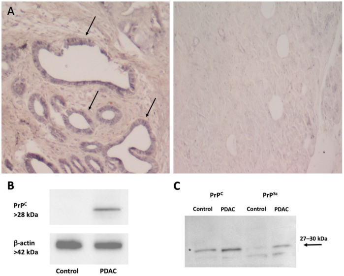
Figure 2.
PrPC expression in human pancreatic tissues. (A) The figure reports representative pictures of PrPC immunohistochemistry of pancreatic ductal adenocarcinoma (PDAC) tissue (left panel) compared with control tissue (right panel). Immunoperoxidase shows PrPC-specific labelling in ductal cells (arrows) from a human tumor sample (left panel). Remarkably, the increase in PrPc expression occurs along with a marked loss of cellular architecture within pancreatic tumoral tissue, which features enlarged and irregularly shaped ducts within abundant extracellular matrix. In contrast, normal human pancreas (right panel) possesses a well-preserved architecture of both acinar cells and ductal system, along with a weak PrPC-staining in the ductal epithelial cells (original magnification 10×). (B) The figure reports a representative immunoblot for PrPC and the house keeping protein β-actin in control and PDAC tissues. (C) The figure reports representative western blotting comparing scrapie prion protein (PrPSc) and PrPC (with or without proteinase K) in control and PDAC tissues as measured in Table 1. Images were obtained by an author of the manuscript (M.A. Giambelluca). Original western blot images (Figures S1 and S2) and densitometry readings (Tables S1 and S2) were provided in Supplemental Materials.

