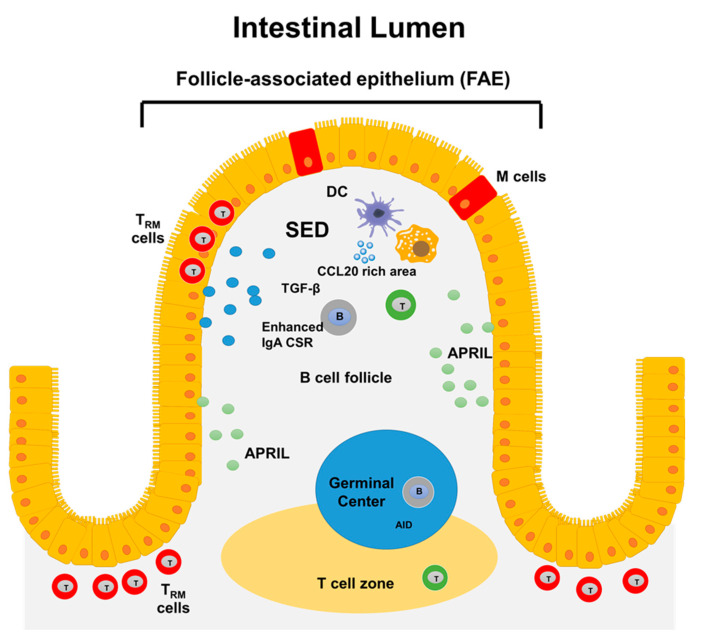
Figure 1.
Structure of GALT. In the gut, antigens are sampled by follicle-associated epithelium (FAE) which contain microfold (M) cells. Next to M cells the sub-epithelial dome (SED) region is located. This region hosts multiple cell types, including different subsets of dendritic cells (DCs), macrophages, neutrophils, B, and T cells. Several factors that favor PCs survival (e.g., APRIL) and CSR to IgA (e.g., TGF-β) are present in GALT. The GALT also contains germinal centers associated with T-cell zones for induction of immune responses at the local level. Finally, the human gut mucosa contains tissue-resident memory T cells, which show evidence of previous antigen encounter and appear to be long-lived.

