Figure 1.
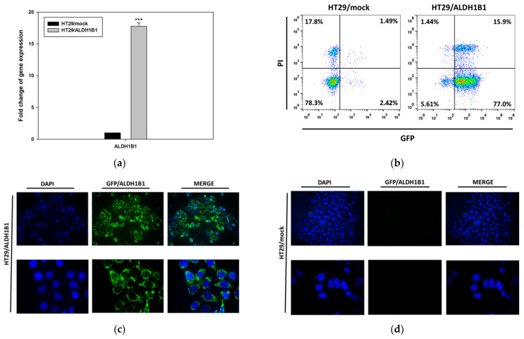
Expression of ALDH1B1 in the HT29 isogenic cell line pair. (a) ALDH1B1 gene expression levels detected by real-time PCR in HT29/mock and HT29/ALDH1B1 cells. (b) Evaluation of GFP+ cells (green) in HT29/mock and HT29/ALDH1B1 by flow cytometry analysis. Nuclei were stained with propidium iodide (PI) (red). Fluorescence microscopy for detecting the GFP+ cells (green) in HT29/mock (c) and HT29/ALDH1B1 (d) cells (upper panel at 40× magnification and lower panel at 100× magnification). Nuclei were stained with DAPI (4′,6-diamino-2-phenylindole) (blue). Results are expressed as mean ± SD of three independent experiments. *** p < 0.001.

