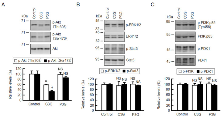
Figure 4.
Effects of anthocyanins on intracellular signaling pathways. Cells were incubated in the absence (control) and presence of 50 µM anthocyanins for 1 h. (A) Cell lysates were immunoblotted with anti-p-Akt (Thr308), anti-p-Akt (ser473), and anti-Akt antibodies. (B) Cell lysates were immunoblotted with anti-p-ERK1/2, anti-ERK1/2, anti-p-Stat3, and anti-Stat3 antibodies. (C) Cell lysates were immunoblotted with anti-p-PI3K p85 (Tyr458), and anti-p-PI3K p85, anti-p-PDK1, and anti-PDK1 antibodies. The protein levels were represented as a percentage relative to control. n = 4. * p < 0.05 and NS p > 0.05 compared with control.

