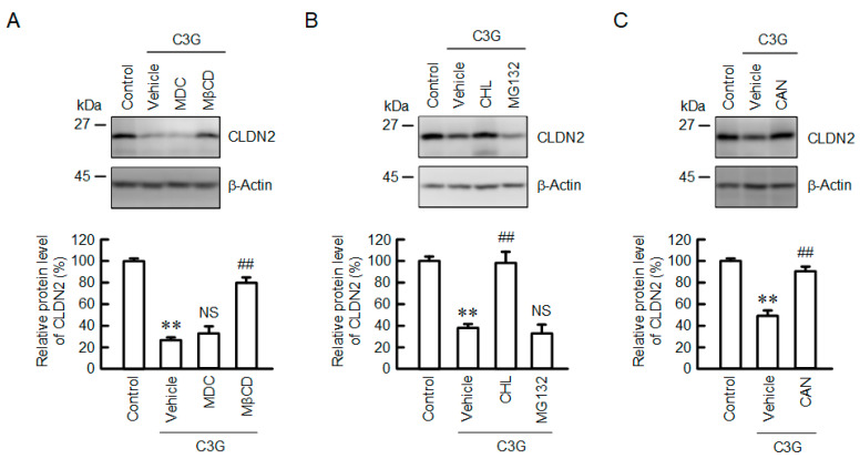
Figure 6.
Effect of C3G on the degradation and endocytosis of the CLDN2 protein. (A) Cells were incubated in the absence (control) and presence of 50 µM C3G, 5 µM MDC, or 10 µM MβCD for 24 h. (B) Cells were incubated in the absence (control) and presence of 50 µM C3G, 20 µM CHL, or 5 µM MG132 for 24 h. (C) Cells were incubated in the absence (control) and presence of 50 µM C3G for 24 h. CAN (0.5 µM) was added into the medium 9 h before collection. Cell lysates were immunoblotted with anti-CLDN2 and anti-β-actin antibodies. The protein levels of CLDN2 are represented as a percentage relative to control. n = 3–5. ** p < 0.01 compared with control. ## p < 0.01, and NS p > 0.05 compared with vehicle.

