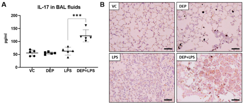
Figure 6.
Protein levels of (A) IL-17 in the bronchoalveolar (BAL) fluid and (B) representative images of immunohistochemistry (IHC) for IL-17 (brown color) in the lung tissue sections from mice in the VC, DEP, LPS, and DEP pre-exposed and LPS-instilled (DEP+LPS) groups. Scale bars = 200 nm. Data represent means ± SD (n = 5 per group) *** p < 0.001 vs. LPS group.

