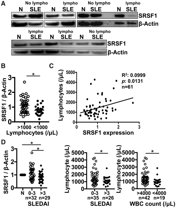
Fig. 1.
Low SRSF1 levels correlate with lymphopenia in patients with SLE
Peripheral blood T cells were isolated from patients with SLE (n = 61) and healthy control individuals (n = 44). Total protein was immunoblotted for SRSF1 and β-actin. (A) Data are from one representative of eight independent experiments. (B) Densitometric quantitation of Western blots was performed and SRSF1 normalized to β-actin. Relative SRSF1 expression in SLE patients was normalized to matched healthy controls. (C) Graph shows linear correlation between relative SRSF1 protein levels and peripheral blood lymphocyte counts (n = 61). (D) Graphs show associations of relative SRSF1 with SLEDAI and lymphocyte counts with SLEDAI or WBC counts [B, D (middle and right): unpaired t test; D (left): one-way analysis of variance with Tukey’s correction; C: single linear regression, *P < 0.05].

