Figure 1.
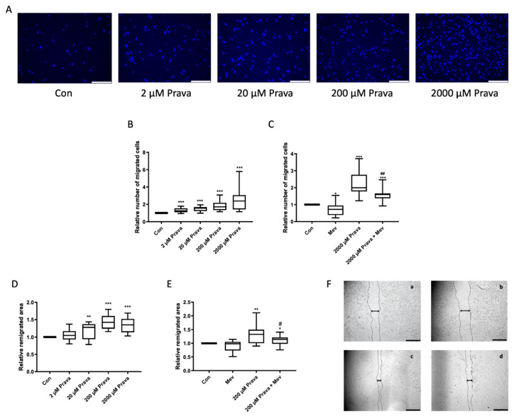
Pravastatin increased endothelial colony-forming cells’ (ECFCs’) migration. (A) Representative images of 4′,6-diamidino-2-phenylindole (DAPI)-stained migrated ECFCs treated with pravastatin (2, 20, 200 and 2000 µM) in a chemotaxis assay, scale bar 150 µm. (B) ECFCs showed a significantly higher directional migration in the presence of pravastatin after 4 h. (C) Mevalonate (200 µM) decreased directional migration and, in combination with pravastatin (2000 µM), significantly decreased the pravastatin induced increase in directional migration. Further representative images can be found in Supplemental Figure S1. Numbers of DAPI-stained migrated cells on the lower side of the membranes were counted in each picture, n = 15–20. (D) Pravastatin treatment (2, 20, 200 and 2000 µM) of ECFCs enhanced wound closure assessed as remigrated area after 18 h compared to control. (E) Mevalonate (200 µM) alone had no significant effect on ECFCs’ migration, but its addition to pravastatin (200 µM) reduced the pravastatin effect significantly. (F) Representative images of monolayers with scratch wounds at 18 h of incubation with culture medium only (a), 200 µM mevalonate (b), 200 µM pravastatin (c) and combination of 200 µM pravastatin and 200 µM mevalonate (d), scale bar 1000 µm. Cell-free area after 18 h was subtracted from cell-free area at start to calculate remigrated area. n = 14–16. Con, control; Prava, pravastatin; Mev, mevalonate. * p < 0.05, ** p < 0.01, *** p < 0.001 compared to control, # p < 0.05 compared to 200 µM pravastatin, ## p < 0.01 compared to 2000 µM pravastatin; (B–E) control group set as 1.

