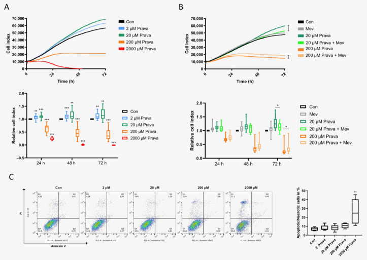
Figure 3.
Pravastatin had a biphasic impact on ECFC proliferation. (A) Overlay of growth curves of ECFCs treated with pravastatin (2 µM, 20 µM, 200 µM or 2000 µM). ECFC proliferation was significantly increased after treatment with 2 µM or 20 µM pravastatin, but significantly decreased after treatment with 200 µM or 2000 µM pravastatin after 24 h, 48 h and 72 h. (B) Overlay of growth curves of ECFCs treated with 20 µM or 200 µM pravastatin in the presence or absence of 200 µM mevalonate. Mevalonate reduced the proliferative effect of 20 µM pravastatin and lessened the antiproliferative effect of 200 µM pravastatin significantly after 72 h. n = 15–20; control group set as 1. (C) High dose pravastatin led to apoptosis. Representative measurement of apoptosis and necrosis in ECFCs after 48 h treatment with pravastatin at 2 µM, 20 µM, 200 µM or 2000 µM. Viable cells are located in the lower left field (Annexin V neg./PI neg). Pravastatin (2000 µM) caused a significantly higher rate of non-viable cells. n = 5. Con, control; Prava, pravastatin; Mev, mevalonate. * p < 0.05, ** p < 0.01, *** p < 0.001.

