Figure 3.
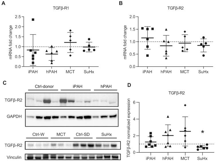
The expression of transforming growth factor-β receptors (TGFβR): (A) the mRNA expression of TGFβR1 analyzed by qPCR; (B) the mRNA expression of TGFβR2 analyzed by qPCR. n(iPAH) = 6, n(hPAH) = 6, n(MCT) = 5, n(SuHx) = 5; (C) representative image of Western blot analysis in whole lung lysates; (D) quantification data of Western blot analysis normalized to vinculin. n(iPAH) = 7, n(hPAH) = 7, n(MCT) = 6, n(SuHx) = 6. Quantification in each group is the fold change corrected by its own control group. The control group of iPAH and hPAH: donors without PAH. The control group of MCT: healthy Wistar rats. The control group of SuHx: healthy Sprague Dawley rats. iPAH: idiopathic pulmonary arterial hypertension, hPAH: hereditary pulmonary arterial hypertension, Ctrl: control, MCT: monocrotaline rats, SuHx: sugen–hypoxia rats. * p < 0.05, compared to its respective control group.

