FIGURE 2.

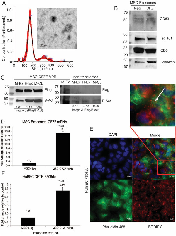
MSC Exosome mediated delivery of Zinc Finger Protein Activator increases the expression of CFTR. (A) TEM micrograph of exosomes isolated from the culture medium of MSC‐CFZF‐VPR transfected cells. Exosomes were measured by using Nanosight NS 300 system in the supernatant from cultures cells. The histogram represents particle size distribution. (B) Western blot analysis for exosome markers in CFZF‐VPR transfected MSC (CFZF) and non‐transfected MSCs (Neg) derived exosomes. (C) Western blot of FLAG‐tagged CFZF‐VPR protein enriched in exosomes from left: MSC‐CFZF‐VPR or right: non‐transfected MSC‐exosomes (M‐Ex), HEK293‐exosomes (H‐Ex) and corresponding MSC cell lysate (M‐CL) samples, respectively. The Image J values from the Flag‐tag (Flag) relative to Beta Actin (B‐Act) are shown below the blot. (D) Evaluation by qRT‐PCR of mRNA expression of exosomes from CFZF‐VPR/Cx43‐transfected MSCs. The results from triplicate exosomes collected from three different CFZF‐VPR/Cx43 transfected MSCs are shown. (E) Light microscopy immunofluorescence images of HuBECs uptake of MSCs‐CFZF‐VPR exosomes labelled with BODIPY TR ceramide (red), Nuclei (Blue), Actin (Green). Scale bar, 10 μm. (F) CFTR expression was determined by qRT‐PCR following treatment with MSC exosomes directed to the CFTR promoter (MSC‐CFZF‐VPR) or Control (MSC‐Neg) in HuBECs. For E and F experiments were performed in triplicate with 10e+03 HuBECs treated with 5e+10 exosomes. Experiment shown the standard error of the means and p values from a paired two‐sided T‐test, *P = 0.01
