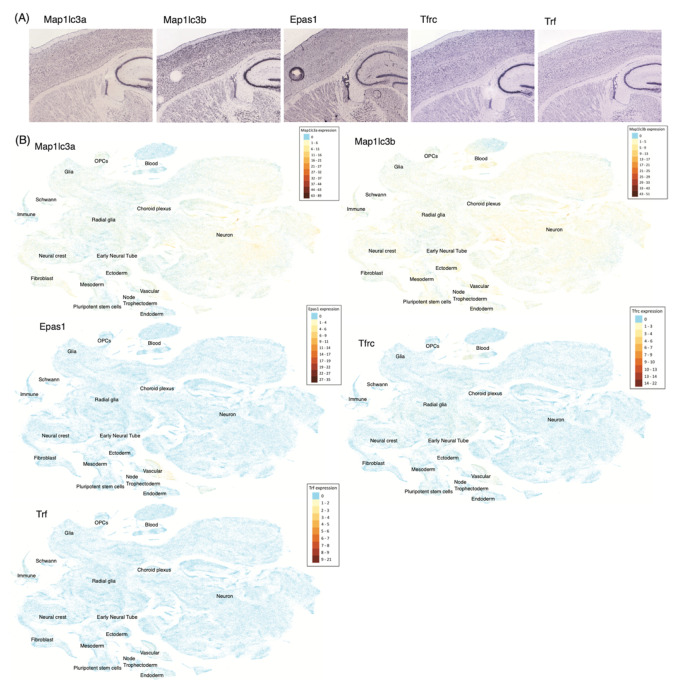
Figure 4.
Ferroptosis gene expressions in the mouse brain. (A) Results of in situ hybridization (ISH) experiments localizing FR gene expressions in the cortices of the P56 mouse brain of Allen Brain ISH Atlas [137]. (B) t-SNE plot of cells expressing FR genes across the developing mouse brain, using the UCSC Cell Browser and the single cell RNA-sequencing data of Manno et al. [138]. Cell types are annotated inside the plots.

