Figure 5.
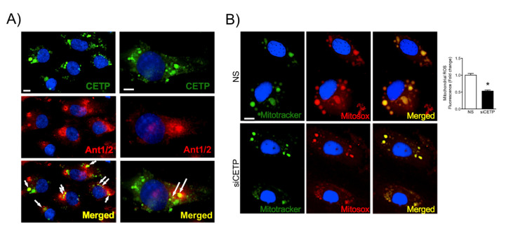
CETP co-localizes partially with mitochondria and increases mitochondrial superoxide production. (A) Representative images of HAECs showing CETP in green, mitochondrial carrier protein adenine nucleotide translocator (Ant1/2) in red, and DAPI in blue (nuclear dye). Arrows indicate co-localization of CETP and ANT1/2 (yellow). Magnification 10X left and 20X right. Scale bar, 10 μm. (B) Representative images of HAEC after CETP silencing and staining with MitoSOX Red (mitochondrial superoxide indicator), Mitotracker Green (mitochondrial marker), and DAPI. Magnification is 10X and x2 zoom. Scale bar, 10 μm. Quantification of co-localization of Mitotracker and Mitosox (n = 5, * p < 0.001). HAECs, human aortic endothelial cells. CETP, cholesteryl ester transfer protein. NS, non-silenced. siCETP, silenced CETP.

