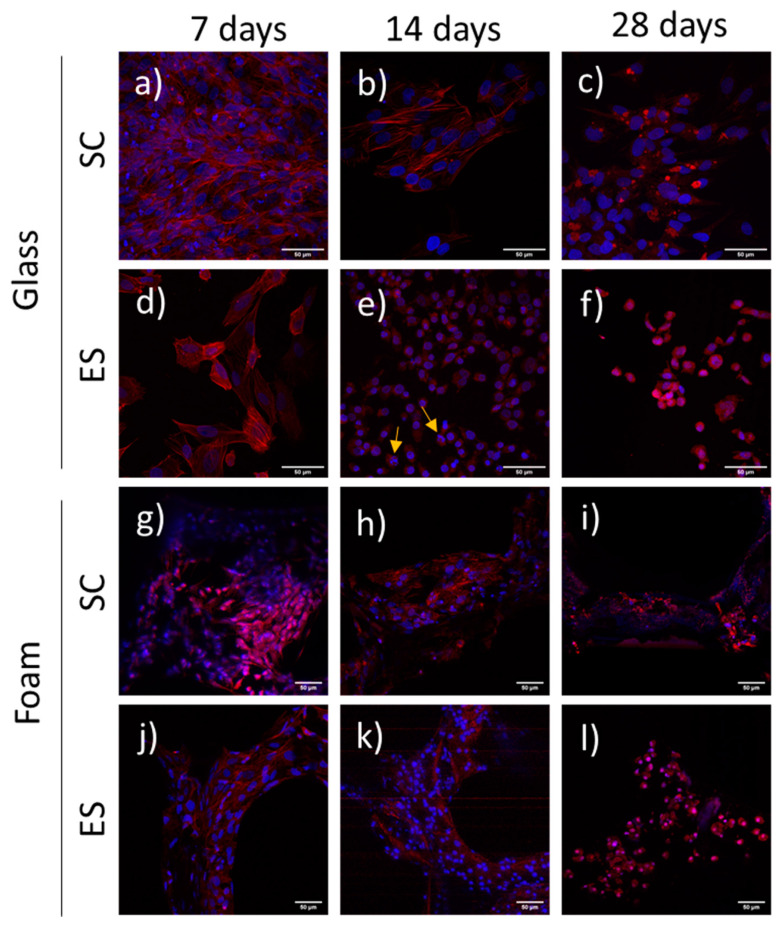
Figure 4.
Actin distribution in human Saos-2 cells cultured for 7, 14 and 28 days under standard conditions (SC) or electrical stimulation (ES) on either glass coverslips (a–f) or Pd-coated PU foams (g–l). Stress fibers (red) and nuclei (blue) can be observed. Yellow arrows point to apoptotic nuclei. Pd-coated PU foams images (g–l) were obtained at less augmentation and digitally augmented due to the nature of the foam material.

